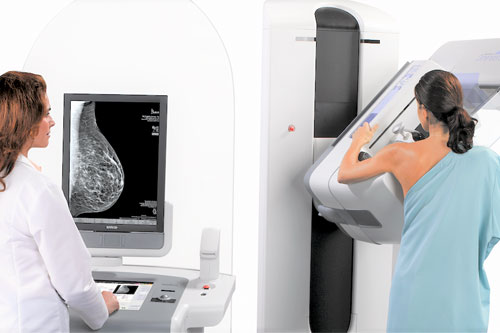
Why 3D Mammography Protects Better Against Breast Cancer

Why 3D Mammography Protects Better Against Breast Cancer
Dr. Pushpinder Singh Khera, HOD, Dept of Radiology
AIIMS, Jodhpur
 Is 3D mammogram better than a 2D? A question asked by many women. Many studies say yes – let’s explore why!
First, what is the difference between 2D and 3D Mammography? In simple words:
- 2D mammography is the older technology. It takes only two images – one from top and the other from the side of the breast, for the doctor to examine. Thus, it may miss overlapping tumors between the tissues, if any.
- 3D mammography exam is the latest technology. It takes many images from different angles, and provides clearer, more detailed pictures. This helps doctors to see better through the breast tissue and discover any tiny tumors that might be “hiding”, which were not visible on 2D mammogram, and on other early screening modalities such as sonography (ultrasound).
Studies have shown that 3D mammography significantly helps in early breast cancer detection, enabling the detection of more cancer cells[1], and significantly reducing recall rates[2] for any additional tests.
Early detection is key for survival and complete cure in breast cancer because there is no way to prevent it. 3D mammography exams help eliminate detection challenges associated with 2D, by using innovative technology designed to produce clear images of the breast tissue, layer by layer[3].
3D mammography is especially useful in high-risk women, i.e., including those with:
- Changes or lumps in the breasts
- A family history of breast or ovarian cancer[4]
- Dense breast tissue (nearly half of all women above the age of 40 have dense breasts)[5]
- A previous diagnosis of breast disease
The 3D Mammography exams detect 20–65% more invasive breast cancers than 2D alone, with an average increase of 41%.
This means two simple things: Earlier detection than ever before, and Less anxiety about unnecessary further testing[6]. In most cases, especially if you routinely examine yourself, it will be good news that your doctor will share that ‘all is well’.
What action can you take today?
If you are eligible for regular breast screening either by age (40 years and above), or due to high-risk factors as above (in which case before 40 years), ask your doctor about an annual 3D Mammography plan – a simple ritual that can save your life!

Dr. Pushpinder Singh Khera,
HOD, Dept of Radiology, AIIMS, Jodhpur
- Skaane, Per, et al. “Comparison of digital mammography alone and digital mammography plus tomosynthesis in a population-based screening program.” Radiology 267.1 (2013): 47-56. https://pubs.rsna.org/doi/full/10.1148/radiol.12121373
- Rose SL, Tidwell AL, Bujnoch LJ, Kushwaha AC, Nordmann AS, Sexton R Jr. Implementation of breast tomosynthesis in a routine screening practice: an observational study. AJR Am J Roentgenol. 2013 Jun;200(6):1401-8. doi: 10.2214/AJR.12.9672. PMID: 23701081. https://pubmed.ncbi.nlm.nih.gov/23701081/
- Friedewald SM, Rafferty EA, Rose SL, Durand MA, Plecha DM, Greenberg JS, Hayes MK, Copit DS, Carlson KL, Cink TM, Barke LD, Greer LN, Miller DP, Conant EF. Breast cancer screening using tomosynthesis in combination with digital mammography. JAMA. 2014 Jun 25;311(24):2499-507. doi: 10.1001/jama.2014.6095. PMID: 25058084. https://pubmed.ncbi.nlm.nih.gov/25058084/
- https://www.canceraustralia.gov.au/sites/default/files/publications/breast-cancer-risk-factors-review-evidence/pdf/rfrw-breast-cancer-risk-factors-a-review-of-the-evidence_1.15.pdf
- https://www.cancer.gov/types/breast/breast-changes/dense-breasts
- Zuley ML, Bandos AI, Ganott MA, Sumkin JH, Kelly AE, Catullo VJ, Rathfon GY, Lu AH, Gur D. Digital breast tomosynthesis versus supplemental diagnostic mammographic views for evaluation of noncalcified breast lesions. Radiology. 2013 Jan;266(1):89-95. doi: 10.1148/radiol.12120552. Epub 2012 Nov 9. PMID: 23143023; PMCID: PMC3528971. https://pubmed.ncbi.nlm.nih.gov/23143023/
